How to Recognise Skin Cancer

The purpose of this guide is to help you identify skin cancer which you might otherwise have missed. The purpose is not to allow you to rule out skin cancer, as this can be very difficult even for a dermatologist. If you are unsure about a skin lesion, or if a lesion is new or changing, it is essential to see a dermatologist.
Skin cancer is the most common cancer in the world, yet with early detection it is often highly treatable. In most cases it can be recognised, yet many people have little idea of what to look for. This guide equips you with the knowledge to better recognise potentially suspicious skin lesions - and you can practise with my skin cancer recognition quiz.
Contents
What Does Skin Cancer Look Like?
Skin cancer can have many different appearances, making it challenging to identify. It is important to be aware of these different presentations. This page gives you examples of common skin cancers and harmless skin lesions.
Changing Mole
An existing mole that changes in size, shape, colour, or develops new symptoms like itching, tenderness, or bleeding. You should see a dermatologist urgently if you notice any change in the appearance of a mole.
New Skin Lesion
A completely new spot, mole, or growth that appears on your skin, particularly if it is asymmetric, has irregular borders, varied colour, or is growing.
Non-Healing Lesion
A sore, cut, or scab that does not heal within a few weeks, or repeatedly heals and breaks open again. This can be a sign of certain types of skin cancer, such as squamous cell carcinoma and basal cell carcinoma.
New Lump or Nodule
A new lump or bump that appears on your skin, which may be skin-coloured, red, or translucent. This can be a sign of squamous cell carcinoma or basal cell carcinoma.
Examples of Skin Lesions
Melanoma
Melanoma is the most dangerous type of skin cancer due to its potential to spread if not caught early. It can develop from an existing mole or appear as a completely new dark lesion on the skin. The following "ABCDE" signs can help you to identify moles that are more concerning:
- Asymmetry: One half of the mole does not match the other half. Imagine drawing a line through the middle - if the two sides look different, it's a warning sign.
- Border: The edges of the mole are irregular, notched, blurred, or ragged. Benign moles usually have smooth, well-defined borders.
- Colour: The colour is uneven, with shades of brown, black, red, white, or blue. A harmless mole typically has one consistent shade of brown.
- Diameter: The mole is larger than 6mm (about the size of a pencil eraser), though melanomas can sometimes be smaller.
- Evolution: Any change in size, shape, colour, elevation, or new symptoms like itching, tenderness, or bleeding.
Here are examples of melanoma:
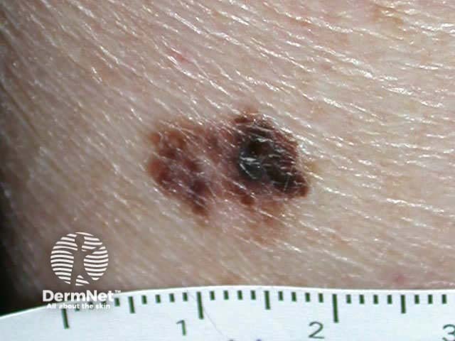
Example of a melanoma demonstrating asymmetry and irregular borders. Image sourced from DermNet
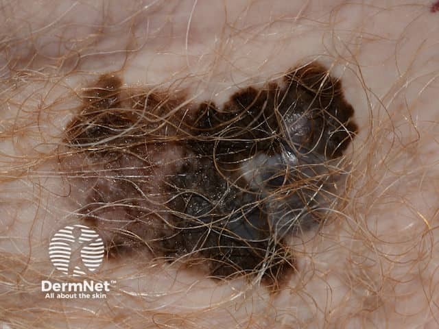
Another example of a melanoma, showing dark brown, light brown and whitish colours with an irregular border. Image sourced from DermNet
Squamous Cell Carcinoma (SCC)
SCC most often appears on sun-exposed areas like the face, ears, lips, and hands but can appear at any site on the body. While less aggressive than melanoma, SCC can be locally invasive and, in some cases, metastasise (spread to the lymph nodes or distant sites in the body) if untreated.
- Can present as a firm, red nodule or bump that grows over time.
- May have a scaly or crusted surface, sometimes resembling a wart.
- Can look like a persistent sore that doesn't heal, or that heals and then re-opens.
- May bleed easily when bumped or scratched.
- Can sometimes be tender or painful to the touch.
Examples of squamous cell carcinoma:
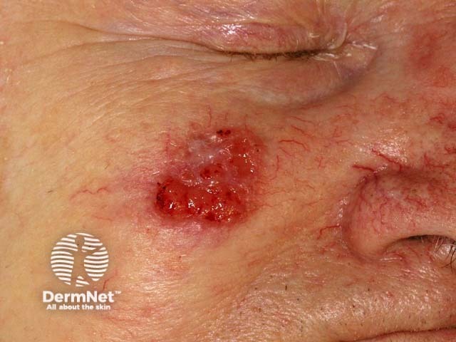
A reddish non-healing lesion that can scale and crust. Image sourced from DermNet
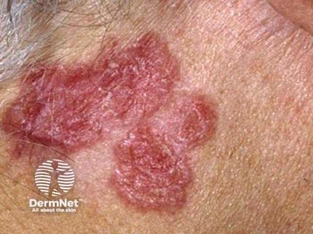
A flat scaly lesion that slowly enlarges with time. Image sourced from DermNet
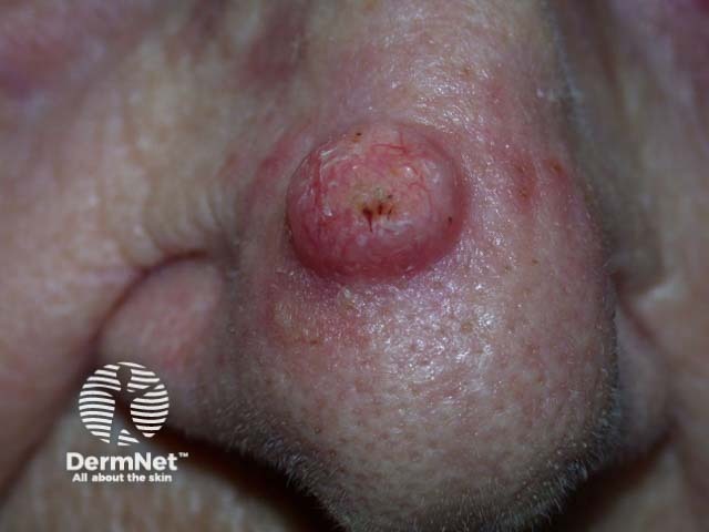
A round lump with central crusting. Image sourced from DermNet
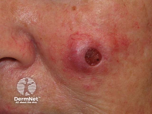
Another round lesion with a central non-healing, crusted area. Image sourced from DermNet
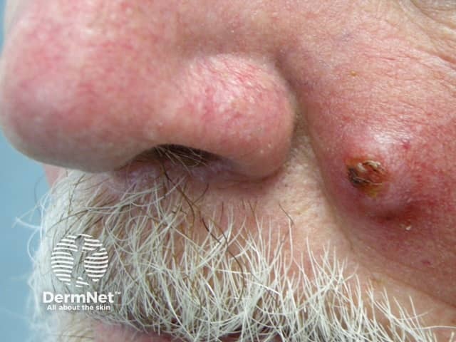
A lump with crusting. Image sourced from DermNet
Basal Cell Carcinoma (BCC)
BCC is the most common type of skin cancer. It grows slowly and rarely spreads to other parts of the body, but it can be locally destructive if not treated, invading surrounding tissues.
- Can look like a pearly or waxy bump, often with a translucent appearance.
- May appear as a skin-coloured or brown scar-like lesion, which can sometimes be subtle and easily missed.
- Often has visible fine blood vessels (telangiectasias) within it, giving it a reddish colour.
- Can present as a persistent, non-healing sore that bleeds or crusts, and may heal and recur.
- Sometimes appears as a red, irritated patch.
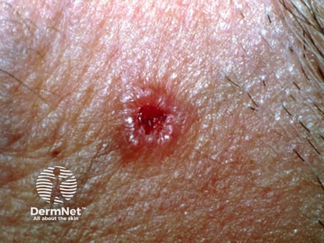
A typical basal cell carcinoma with a pearly appearance and central ulceration. Image sourced from DermNet
Seborrhoeic Keratosis
These are extremely common, benign (non-cancerous) skin growths that usually appear in middle-aged and older adults. While they are harmless, their varied appearance can sometimes cause concern, leading them to be mistaken for skin cancer. They can appear almost anywhere on the body.
- Appear as waxy, "stuck-on" looking growths, as if someone pasted them onto the skin.
- Can be skin-coloured, brown, black, or light tan in colour.
- Often have a slightly raised, rough, or bumpy surface, which can feel greasy or crumbly.
- Vary greatly in size, from tiny dots to more than an inch across, and can appear anywhere on the skin, including the face, chest, back, and scalp.
A seborrhoeic keratosis often has a "pasted on" appearance:
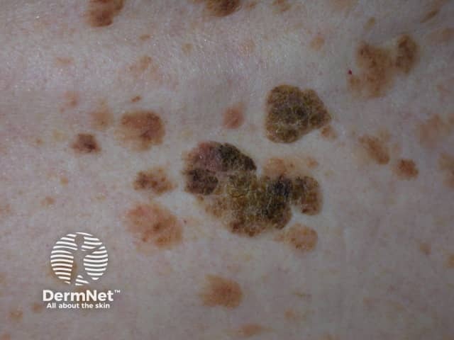
A cluster of seborrhoeic keratoses. Image sourced from DermNet
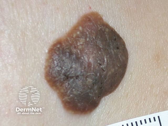
A seborrhoeic keratosis with a uniform colour and well delineated margin. Image sourced from DermNet
Harmless Mole (Nevus)
Most moles are harmless and have the following features:
- Usually symmetrical, meaning if you draw a line through the middle, both halves match.
- Even, smooth borders that are clearly defined against the surrounding skin.
- Single, consistent shade of brown, though they can vary from light tan to dark brown.
- Often smaller than 6mm in diameter, but can be larger.
- Stable over time, meaning they do not change in size, shape, colour, or symptoms.
Here are some common benign moles:
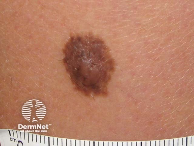
An example of a benign mole with uniform colour and smooth borders. Image sourced from DermNet
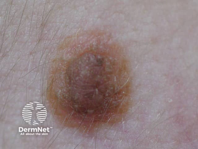
A harmless "compound" naevus (mole) with a central raised area but a symmetrical appearance, uniform colour and smooth borders. Image sourced from DermNet
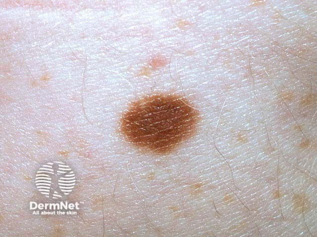
A flat, brown mole with uniform colour and smooth borders. Image sourced from DermNet
Lentigo
Lentigines are small, pigmented spots on the skin. They are usually benign, but some forms (like solar lentigines or "sun spots" / "age spots") can sometimes be difficult to distinguish from early melanoma, especially if they are irregular. They are caused by increased pigment production rather than an increase in pigment cells.
- Appear as flat, uniformly brown or black spots on the skin.
- Often have a uniform colour and regular border, though solar lentigines can sometimes be slightly irregular.
- Commonly found on sun-exposed areas such as the face, hands, shoulders, and arms.
- Do not significantly change over time in terms of size or colour.
These images show typical lentigines:
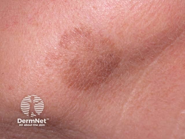
A flat, brown solar lentigo, also known as a sun spot or age spot. Image sourced from DermNet
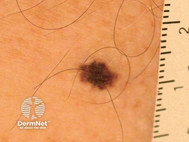
A darker lentigo with a symmetrical pattern. This can be harder to distinguish from a worrying mole and may need to be checked by a dermatologist. Image sourced from DermNet
Haemangioma
Haemangiomas, also known as cherry angiomas, are benign growths made up of small blood vessels. They are extremely common, especially after the age of 30, and usually cause no symptoms. They are not related to sun exposure or skin cancer.
- Can appear as bright red, cherry-like bumps on the skin.
- May be flat, slightly raised, or dome-shaped.
- Often blanch (turn pale) when pressed, though this can be subtle with smaller lesions.
- Usually appear in adulthood and can increase in number with age, varying in size from pinpoint to several millimetres.
- Most commonly found on the trunk, arms, and shoulders.
Examples of a haemangioma:
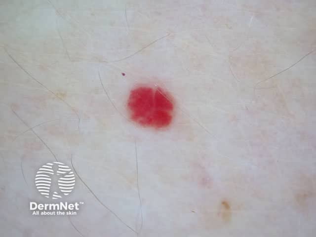
A classic cherry angioma (haemangioma), a common benign vascular lesion with a reddish colour and smooth border. Image sourced from DermNet
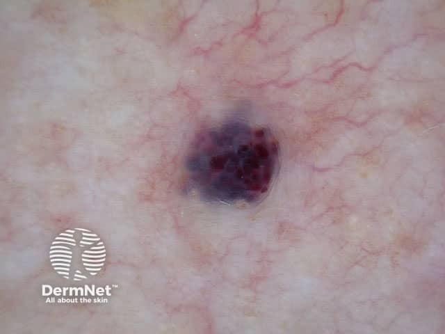
Another angioma appearing as a cluster of purple globules. Image sourced from DermNet
Test yourself: reading about skin cancer is one thing - recognising it is another. Try the skin cancer recognition quiz: real lesion photographs, and instant feedback on whether concern was warranted.
How to Check Your Skin
Regular self-examination is crucial for the early detection of skin cancer.
Consistent Self-Monitoring with Photos
You should regularly monitor your own skin at home. Take full-body photos, and close-up photos of specific moles, every couple of months. Compare these on a computer screen looking for any change in the appearance of existing lesions or any new lesions. Pay close attention to areas that are difficult to see, like your back, scalp, and buttocks - if there is someone at home ask them to help you to photograph or examine these sites.
Full Skin Check with a Dermatologist
It can be very difficult to know whether to worry about a particular skin lesion. A good option is to start by having a professional dermatologist perform a thorough full-body skin examination. Once you know that existing skin lesions are harmless, you can focus on looking for any change in appearance.
Get to Know Your Skin
Become familiar with your moles, freckles, and other skin lesions. Understanding what is "normal" for your skin will help you quickly identify anything new, changing, or unusual.
If in Doubt, Check it Out
If you notice any lesion that is new, changing in any way (size, shape, colour, sensation like itching or bleeding), or simply makes you feel uneasy, do not hesitate to consult a dermatologist.
When to See a Doctor
If you notice any new skin lesions or see any changes in existing ones, consult a dermatologist or your primary care doctor without delay. Any lesion that is growing, bleeding, itching, or changing in appearance should be checked. Early detection significantly increases the chances of successful treatment for all types of skin cancer.
Dr Lynch offers skin cancer checks and mole checks at his Central London clinics.